Important Dates
- Abstract Deadline: Wednesday, March 25, 2026
- Early Bird Registration: Saturday, February 28, 2026
- Regular Registration Deadline: Monday, March 30, 2026
- Conference Date: Thursday, April 30, 2026
Aims & Scope
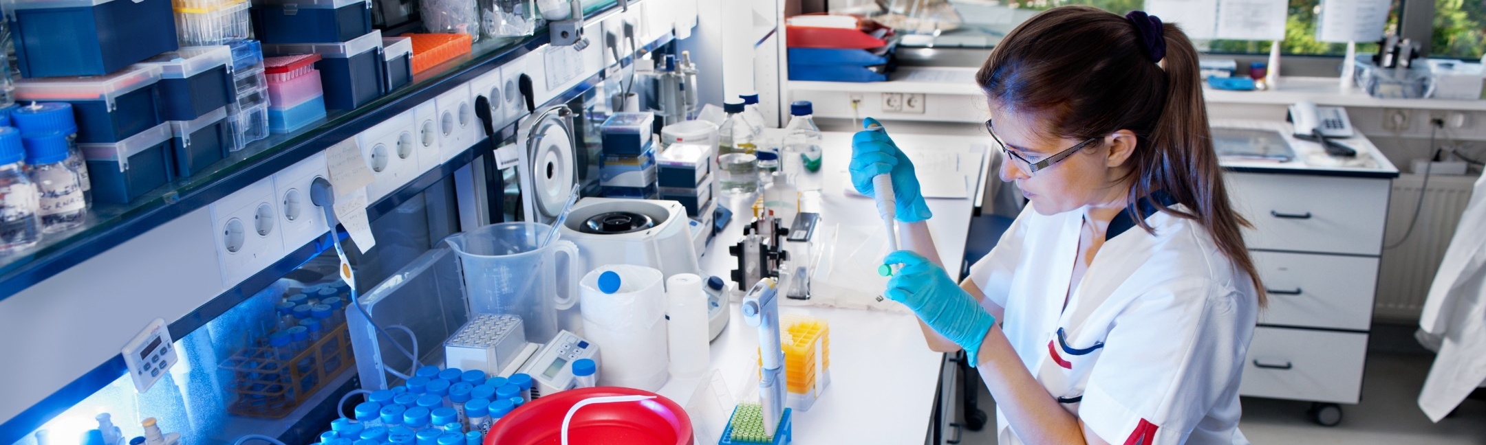
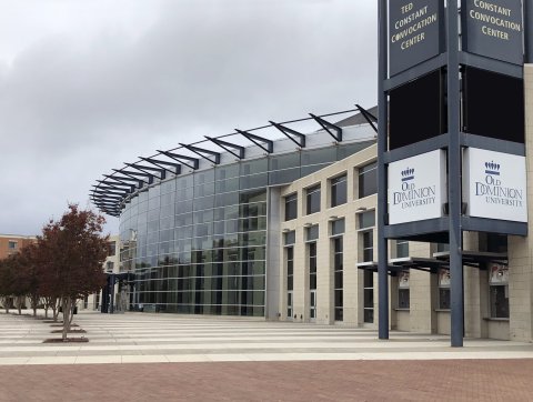
NanoBioTech's Biomedical Innovation and Integrated Health Systems (BIIHS) 2026 Conference will be held on April 30, 2026, at the ODU Ted Constant Convocation Center. The event will bring together leading experts, industry partners, students, and staff to discuss cutting-edge developments in biomedical research and integrated health systems.
The conference will showcase advances in diagnostics, therapeutics, biomaterials, nanotechnology, environmental health, education, and the materials and computational research that enable these technologies.
The conference format includes plenary talks from invited speakers of international renown and oral presentations describing new research selected based on their originality and scientific quality.
- A highly focused 1-day conference, dual-track oral sessions
- Oral presentations will precede the poster sessions
- Presentation of each poster
- Awards for poster presentations
Speakers
Dr. Reza Ghodssi
Title: Revolutionizing Gut Health: Ingestible Devices and Technologies for Disease Diagnosis and Treatment
Dr. Reza Ghodssi is a Distinguished University Professor, the Herbert Rabin Distinguished Chair in Engineering, and a Distinguished Scholar Teacher at the University of Maryland (UMD). Dr. Ghodssi is Director of the MEMS Sensors and Actuators Lab (MSAL) in the Department of Electrical and Computer Engineering (ECE) and the Institute for Systems Research (ISR) and the Inaugural Executive Director of Research and Innovation of the MATRIX Lab at UMD. Dr. Ghodssi's research interests are in the design and development of micro-, nano-, and bio-devices and systems for chemical and biological sensors and actuators, and small-scale energy conversion and harvesting for healthcare applications. Dr. Ghodssi is President of the Transducer Research Foundation (TRF), a Fellow of IEEE, AVS, and ASME, has 178 journal publications and 378 refereed conference papers, and is the co-editor of the MEMS Materials and Processes Handbook published in 2011. Dr. Ghodssi served as chair and technical program chair of several national and international MEMS conferences, including the 2022 and 2020 Hilton Head Workshops. Dr. Ghodssi was the lead organizer and chair of the inaugural Denice Denton Emerging Leaders Workshop 2016 held in Madison, Wisconsin, which focused on helping mid-career faculty (women and men) develop knowledge, skills, strategies, and critical networks. He served as an associate editor for the Biomedical Microdevices (BMMD) for 13 years (2008-2021) and served as an associate editor for the Journal of Microelectromechanical Systems (JMEMS) for twelve years (2008-2020). He has ten U.S. patents issued, nine U.S. patents published, and another seven pending. Dr. Ghodssi received several awards and recognitions, including the 2024 American Vacuum Society (AVS) Gaede-Langmuir Award.
Abstract:
Gastrointestinal (GI) diseases, including inflammatory bowel disease (IBD) and various forms of cancer, are increasingly prevalent due to a combination of genetic, environmental, and lifestyle factors, impacting more than 40 million people in the US alone. Current medical tools for monitoring and treating GI diseases date back to the 1970s and require invasive procedures. An emerging method for probing the GI tract is the use of minimally invasive devices to monitor, detect, diagnose, and treat, particularly in remote regions of the GI. These devices rely on MEMS and microsystems, which have demonstrated the potential to improve healthcare through compact, low-power, and cost-effective solutions for continuous, real-time monitoring and advanced diagnostics, enabling early disease detection, and out-patient digital healthcare. Microsystems, such as wearable electronics, that interface with the body have been thoroughly explored both academically and commercially. Recently, the application of MEMS and Microsystems to ingestible devices has yielded key technologies to address GI-related diseases. The accessible nature of the GI tract provides a gateway for analyzing bodily processes and reaching specific organ systems for treatment.
My group’s research focuses on the development of ingestible tools for monitoring and treatment of GI and systemic diseases. Our integrated devices featuring embedded electronics and sensors enable analysis of critical biomarkers, like hydrogen sulfide (H2S), neurotransmitters, and tissue permeability, while actuators allow sampling and drug delivery at precise locations in tissue for highly effective on-command treatment. It is our hope that these technologies will not only be integrated into ingestible devices but also prove worthy of addressing these problems, making such monitoring tools more accessible to a larger population around the world. In this talk, I discuss some of the challenges and future opportunities of these integrative micro/nano/bio technologies and systems.
Dr. Azahar Ali
Title: From Nanofabrication to Integrated Health Systems: Reimagining Biosensing Beyond the Lab
Dr. Azahar Ali is a tenure-track Assistant Professor in the School of Animal Sciences at Virginia Tech, where he leads research in biosensor engineering with a focus on precision animal farming and integrated health monitoring. He holds affiliate faculty appointments in Biological Systems Engineering and is affiliated with the Center for Advanced Innovation in Agriculture and the Center for Emerging Zoonotic and Arthropod-Borne Pathogens (CeZAP) at Virginia Tech.
Dr. Ali’s research bridges micro- and nanofabrication, additive manufacturing, and electrochemical sensing to develop deployable biosensors for agricultural, biomedical, and environmental applications. Prior to joining Virginia Tech, he conducted postdoctoral research at Carnegie Mellon University, served as a Visiting Researcher at the Hillman Cancer Center, University of Pittsburgh Medical Center (UPMC), and was a Postdoctoral Researcher at Iowa State University. He earned his Ph.D. in Biomedical Engineering from the Indian Institute of Technology Hyderabad.
Dr. Ali has authored 81 peer-reviewed journal articles, delivered more than two dozen conference presentations, and holds 16 filed patents. His work has been cited nearly 6055 times (h-index 46, i10-index 76). He is the recipient of several honors, including the Distinguished Alumni Award from IIT Hyderabad (2023) and the Virginia Tech By-Example Award (2025), recognizing his contributions to high-impact, translational biosensing technologies.
Abstract:
Modern health systems across human, animal, and environmental domains remain largely reactive, relying on episodic, laboratory-based measurements that provide delayed and incomplete insight into dynamic biological processes. Despite major advances in biosensor sensitivity and analytical performance, most sensing platforms remain optimized for controlled experimental conditions rather than the complex, evolving environments in which real-world health decisions are made. This gap between laboratory innovation and deployable diagnostics continues to limit the real-world impact of biosensing technologies. In this keynote, I will present a unifying vision for reimagining biosensing beyond the lab, positioning advanced manufacturing as a central enabler of translation. I will highlight how lithography-based micro- and nanofabrication, together with scalable additive manufacturing and three-dimensional printing, provide unprecedented design freedom over sensor geometry, architecture, and system integration. These capabilities allow biosensors to be engineered around biological reality, including variability, motion, and continuous interaction, rather than static test conditions. The talk will feature representative case studies illustrating how this manufacturing-driven design philosophy translates across diverse applications. Examples include printed biosensors for SARS-CoV-2 detection, neurochemical sensing of dopamine, and highly sensitive sensing platforms for the early detection of subclinical hypocalcemia and mastitis in dairy cows, where timely intervention is critical but conventional diagnostics remain delayed or episodic. Together, these examples demonstrate how a common fabrication and systems approach can be adapted across human and animal health contexts. The keynote will conclude with a forward-looking perspective on how advanced manufacturing–enabled biosensors, when integrated with edge electronics, machine learning, and system-level analytics, can serve as foundational infrastructure for next-generation integrated health systems. Such systems have the potential to shift health monitoring from episodic testing to continuous insight, and from reactive response to proactive, data-driven decision-making across clinical, agricultural, and environmental domains.
Dr. Netz Arroyo
Title: Insights into the Transport of Molecules Across Body Compartments via Aptamer Sensors
Netzahualcóyotl Arroyo Currás, known as Netz Arroyo, is an Associate Professor of Chemistry at University of North Carolina (UNC) at Chapel Hill. He obtained his Ph.D. in 2014 from The University of Texas at Austin under the guidance of Allen J. Bard. From 2015 to 2018 he completed postdoctoral training under Dr. Kevin W. Plaxco at University of California Santa Barbara. He became Assistant Professor of Pharmacology and Molecular Sciences at Johns Hopkins University School of Medicine (JHUSOM) in 2019 and was promoted to the rank of Associate Professor in 2023. In 2024, he was invited to move his research program to UNC, where he currently works since 2025. His laboratory pursues the development of electrochemical biosensors for continuous molecular monitoring in the body, and for clinical diagnostics. He was named a Rising Star in Sensing by the journal ACS Sensors in 2020, and in 2023 his research was highlighted as highly impactful by the journal Langmuir, both publications of the American Chemical Society. Dr. Arroyo serves as Interim Editor-in-Chief of the newly launched journal Sensors Plus by The Electrochemical Society. He is funded by the NIH, AFRL, several corporate sponsorships and by private foundations. In his free time, he enjoys playing, dancing, and eating ice cream with his daughters in the lovely city of Chapel Hill, NC.
Abstract:
The Netz Lab at UNC develops electrochemical biosensors and continuous molecular monitoring technologies to enable real-time, personalized health management. Our work centers on electrochemical aptamer-based (E-AB) sensors that use structure-switching receptors for highly selective and reversible detection of clinically relevant biomarkers. By combining advanced surface chemistry, redox reporter optimization, and computational modeling, we design sensors that are stable, miniaturized, and biocompatible for wearable or implantable platforms. Current efforts focus on continuous monitoring of protein biomarkers to assess metabolic health, with additional applications in therapeutic drug monitoring and point-of-care diagnostics. This presentation will highlight our progress in interfacing sensors with the body and addressing questions of molecular transport across tissue compartments. Ultimately, we aim to answer a key question: Which molecules can be measured minimally invasively through the skin while accurately reflecting medically relevant blood concentrations?
Michael Daniele
Title: How Much Data is Enough? From Continuous Wearables and Insterables to Single-Use Patches Biochemical Data
Michael Daniele is a Professor and University Faculty Scholar in the Departments of Electrical and Computer Engineering and the Lampe Joint Department of Biomedical Engineering at North Carolina State University and the University of North Carolina at Chapel Hill. He co-founded the North Carolina Viral Vector Initiative in Research and Learning (NC VVIRAL) and the NC State Institute for Connected Sensor-Systems (IConS), and he is the current Co-Director of the NSF Nanosystems Engineering Research Center for Advanced Self-Powered Systems of Integrated Sensors and Technologies (ASSIST). He joined the faculty in 2015 from the U.S. Naval Research Laboratory, where he was a Jerome and Isabella Karle Distinguished Scholar in Materials Engineering. In 2019, he co-founded DermiSense, Inc., a medical device startup developing blood-free diagnostics, and he currently serves as its Chief Science Officer. Dr. Daniele’s research focuses on engineering materials and microsystems for wearable biosensors, microphysiological systems, and translational biotechnologies that monitor, mimic, or augment biological function.
Abstract:
Wearable technologies have transformed the capture of electrical, optical, and mechanical biosignals, accelerating translation into clinical and consumer devices. Biochemical sensing is now following a similar trajectory, but it spans multiple operating modes that each impose different constraints on sampling, sensor stability, calibration, and user workflow. In this talk, I will present a spectrum of on-body biochemical biosensing approaches, moving from sweat-based wearables, to continuous dermal interstitial fluid (ISF) monitoring, to rapid single-use microneedle-enabled tests designed for near-immediate decision-making. A central theme will be the field’s pivot from sweat to dermal ISF. Sweat is convenient but often suffers from uncertain systemic correlation, contamination risk, and highly variable sampling kinetics. Dermal ISF offers a more stable and clinically relevant biomarker milieu, but requires engineered interfaces to enable safe, low-burden access and repeatable mass transport to the sensor. I will highlight our work on biosensor integration strategies that address these challenges across both continuous wearables and disposable formats. Finally, I will describe an alternative and complementary path: single-use ISF biosensors that leverage microneedle interfaces to deliver rapid, clinically actionable biochemical measurements with a user experience closer to a “test strip” than a durable wearable. Rather than optimizing for multi-day operation, these devices prioritize speed, simplicity, and consistency, enabling high-confidence measurements in minutes for applications such as point-of-care testing, therapeutic monitoring, and decentralized screening. Together, these modes illustrate how thoughtful integration of skin-access interfaces, microfluidics, and low-power bioelectronics can unlock scalable, accessible biochemical sensing for both clinical and consumer health.
Dr. Vamsi Yadavalli
Title: Nature-derived, photo-actuated biomaterials for the fabrication of functional biodevices
Dr. Vamsi Yadavalli is a Professor in the Department of Chemical and Life Science Engineering at Virginia Commonwealth University in Richmond, Virginia, USA. He is currently serving as a Program Director in the Engineering Biology and Health Cluster within the Chemical, Bioengineering, Environmental and Transport Systems (CBET) Division at the National Science Foundation (NSF).
Dr. Yadavalli received his PhD from Penn State University and conducted postdoctoral research at the National Institutes of Health. His group currently works at the interface of materials science, nanotechnology, and biology to understand multiscale behavior from the nano to the macro scales. He has published over 100 papers in top journals including Advanced Materials, Biosensors & Biolectronics, and ACS journals. He holds several US Patents and is a member of the VCU chapter of the National Academy of Inventors. He received a Fulbright Scholar award to visit the University of Trento, Italy, conducting research in the area of silk biomaterials. His research has been supported by the Dreyfus Foundation, NSF, DTRA, and Commonwealth of Virginia grants.
Abstract:
We are at a transitional phase in materials technologies, with a shift from rigid components to soft and flexible systems. Such devices have broad applications for energy and the environment, including electronic skins, soft robotics, tissue engineering, and wearable or implantable sensors for continuous monitoring of bioinformation. Bioinspired and bioderived materials obtained from the silk cocoon provide an exciting pathway towards creating precise and personalized tools, while addressing issues of environmental sustainability and mitigation of electronic waste. Both silk fibroin and silk sericin have a unique palette of properties that make them extremely versatile and viable candidates for soft (bio)electronics in diverse forms.
Silk proteins can serve as both the passive and active components either by themselves or as composites with a diverse multifunctional materials. Composites with organics such as conducting polymers provide added functionality, together with tunable properties, biocompatibility and biodegradability, which provides prospects for sustainable engineering. This talk will discuss work in transitioning to natural precursors and silk-based biocomposites that can be used as wearable sensors and implantable devices, biophotonic elements, energy-storage devices, assistive technologies, and human-machine interfaces. By integrating microfabrication with silk proteins, we show how high resolution, high-fidelity bio devices can be formed in both rigid and flexible formats in two and three dimensions. Such devices can be interfaced with soft tissue to provide two-way communication. The ease of fabrication, biochemical functionalization, biocompatibility, as well as tunable mechanical properties and biodegradation of these biomaterials provide unique possibilities as environmentally sustainable, devices for broad ranging applications.

Dr. Ozlem Dilek
Title: Click-to-Light: Fluorescent Probe Designs for Tracking Oxidative Stress
Dr. Özlem Dilek is a chemical biologist with a broad interdisciplinary background in chemistry and biomedical research. She earned her B.Sc. and M.Sc. degrees in Chemistry from Middle East Technical University in Ankara, Turkey, and received her Ph.D. in Chemistry/Chemical Biology from the State University of New York (SUNY) at Binghamton. She went on to complete postdoctoral training at both Cornell University and SUNY. In 2013, Dr. Dilek joined the one of the private medical schools (Altinbas University) in Istanbul, where she earned tenure. From 2017 to 2023, she held faculty positions at various institutions across Maine, Connecticut, and Washington, D.C., further expanding her academic and research portfolio. Since Fall 2023, Dr. Dilek has been working as an Assistant Professor in the Department of Chemistry and Biochemistry at George Mason University. She also holds a secondary appointment as Adjunct Research Faculty at the Georgetown University School of Medicine. Her research is highly interdisciplinary, integrating organic chemistry, cell biology, and spectroscopy. Her lab focuses on developing innovative chemical biology tools, particularly novel fluorescent probes using bioorthogonal click chemistry. These technologies allow for precise imaging of biomolecules, contributing to a deeper understanding of cellular processes and human disease mechanisms.
Abstract
Click chemistry has emerged as a versatile platform for creating next-generation imaging probes, enabling precise labeling of intracellular structures and real-time tracking of biological pathways through fluorescence microscopy. However, effective imaging still demands probes with high signal-to-noise ratios, rapid reaction kinetics, and tunable photophysical properties—requirements not consistently met by traditional antibodies and fluorophores due to limitations in size, solubility, and photostability. In this work, we introduce a streamlined click-chemistry strategy for engineering small-molecule probes with optimized chemical and spectroscopic performance. These probes span a wide spectral range and are designed for rapid detection of carbonyl species and metal ions relevant to oxidative stress biology. Spectroscopic characterization and fluorescence microscopy confirm that the probes can differentiate breast cancer cells from normal cells under oxidative stress, demonstrating their diagnostic potential. Key photophysical features observed—including pronounced red shifts, large Stokes shifts upon target binding, and metal ion–induced fluorescence activation—underscore their utility for dynamic, real-time cellular imaging. We further integrate our fluorophores into an innovative 3D bioprinting platform that enables continuous, non-destructive optical monitoring of printed tissues. Beyond fundamental imaging capabilities, our probe-based approach enables monitoring of oxidative stress pathways and other dynamic cellular processes. We anticipate that this class of small-molecule probes will not only accelerate the discovery of therapeutics targeting specific biomolecules but also advance next-generation 3D bioprinting methodologies. By enabling minimally invasive, real-time visualization of tissue maturation, our technology offers a transformative tool for evaluating functional integration in regenerative medicine.
Dr. Chris Highley
Title: Designing dynamic properties and porosity in granular biomaterials using electrospun nanofibers in support of tissue engineering and regeneration
Dr. Chris Highley is an Assistant Professor in the Departments of Biomedical Engineering and Chemical Engineering at the University of Virginia. His laboratory works on developing technology for the fabrication of biomaterial and cellular systems to address biomedical challenges in tissue engineering, regenerative medicine, and the design of microphysiological systems. A central aim of the lab’s work is advancing capabilities to build biological constructs that achieve design-based, functional outcomes through precise multiscale control of construct structure and composition. To achieve this his lab maintains a focus on the development and application of dynamic materials that enable these goals and that may find immediate application in addressing medical challenges. He received his B.S.E. in Biomedical Engineering at Duke University and his Ph.D. in Biomedical Engineering at Carnegie Mellon University. He was a postdoctoral researcher at the University of Pennsylvania before beginning his independent career.
Abstract:
In biomaterial systems used in tissue engineering, in regenerative medicine, and in microphysiological systems there is a critical need for materials that permit cellular proliferation, migration, and self-organization processes that result in tissue structures with physiological function. Granular hydrogel materials have potential to address this need, as they have intrinsic permissive properties resulting from microporosity among component microparticles and, when not crosslinked to one another, the potential for individual microparticle grains to move under applied forces. While granular hydrogels are typically formed from spherical particles, the inclusion of hydrogel nanofibers, or the use of fibers alone, within a granular hydrogel presents opportunities to enhance porosities or engineer mechanical properties that are not available in traditional granular or non-granular hydrogels. Hydrogel fibers included with spherical hydrogel microparticles can enable enhanced porosity, where porosity could be defined by a ratio of stable particles to removable particles. Within these systems, long-range fiber-mediated crosslinking can stabilize granular systems that would otherwise degrade by erosion, and these scaffolds support proliferation of cells in vitro, as well as porosity that can be sustained in vivo after minimally invasive delivery to support revascularization. When hydrogel fibers were used as the sole granular component of a granular hydrogel, rheological and extensibility testing showed that, like traditional granular hydrogels, fiber-based hydrogels exhibited a yield stress and can transition between solid- and liquid-like behaviors depending on applied stresses. Unlike traditional particle-based hydrogels, however, they were highly extensible and exhibit rapid stress relaxation that can be controlled through formulation. In extrusion-based applications, such as bioprinting, the long-range entanglements stabilize printed filaments, even absent interfiber crosslinking. Our results indicate that these materials’ permissive properties support dynamic cellular behaviors such as spreading, proliferation, and the development of multicellular networks, presenting new opportunities for designing 3D biomaterials for applications where permissive properties are critical to cellular properties or the emergence of physiological function.
Dr. Daeha Jung
Title: 3D Printing of Functional Materials for Biomedical Applications
Daeha Joung is an Associate Professor of Physics and Associate Director of the Nanoscience Ph.D. Program at Virginia Commonwealth University. He received his Ph.D. in Physics (Nanoscience) from the University of Central Florida and completed postdoctoral training in the Departments of Electrical Engineering and Mechanical Engineering at the University of Minnesota. His research advances interdisciplinary innovation in biomedical physics by integrating principles from physics, materials science, engineering, and biomedicine. Guided by a physics-driven design philosophy, his group develops functional materials and advanced manufacturing strategies for next-generation human–machine interfaces, regenerative medicine platforms, and adaptive bioelectronic systems. He is an NSF CMMI Panel Fellow and serves on the editorial boards of Micromachines and the International Journal of Medical Devices.
Abstract:
Abstract: Biological systems—from molecules and organelles to cells, tissues, and entire organisms—are defined by complex three-dimensional (3D) architectures. Replicating, interfacing with, or sensing biological function therefore requires artificial systems capable of matching this structural and functional complexity across multiple scales. Traditional fabrication approaches, while powerful for planar and two-dimensional networks, are inherently limited in addressing the geometric intricacy and multi-material integration demanded by next-generation biomedical devices. Our strategy leverages extrusion-based multi-material 3D printing as a versatile additive manufacturing platform for constructing hierarchically organized, functional architectures. This approach enables: (i) the design of personalized 3D-printed biomedical devices, (ii) the incorporation of one-part 3D printable nanofiller–polymer composite inks to introduce tunable electrical, mechanical, and magnetic functionalities, and (iii) the integration of biocompatible and functional materials into unified 3D-bioprinted “living” platforms that promote seamless interaction between engineered devices and human tissues. By combining multi-material manufacturing with physics-driven design principles, we enhance the sensitivity, durability, and adaptability of printed systems for biomedical applications. In this talk, I will highlight our recent progress in developing 3D printed functional materials and devices for biomedical applications.
Dr. Nastassja Lewinski
Title: Nanotechnology and new approach methodologies for coral resilience
Nastassja Lewinski is an Associate Professor of Chemical and Life Science Engineering at Virginia Commonwealth University. She joined VCU in 2014 after completing postdoctoral training at the Institute for Work and Health in Lausanne, Switzerland. She earned her Ph.D. in Bioengineering and B.S. in Chemical Engineering from Rice University. Dr. Lewinski’s research focuses on integrating biological and environmental compatibility into the design of engineered nanoparticles and evaluating biological and environmental compatibility of incidental nanoparticles. Projects have ranged from designing nanomedicines for human and coral health and designing cell-based sensors for human and plant health. She has published 50 papers and has directly advised 10 graduate students and 64 undergraduate students.
Abstract
Nanotechnology has been transformative in human healthcare and these learnings can be leveraged to develop new medicines and devices to promote resilience beyond humans. This presentation will discuss research to develop ceria and polylactic-co-glycolic acid based nanomedicines for reef building corals to mitigate bleaching and enhance tissue repair. Because coral toxicology studies are primarily conducted in vivo using small fragments of coral colonies, advances in coral cell culture to enable higher throughput study of coral responses to chemical intervention strategies will also be discussed. Nanotechnology and new approach methodologies can expand the available toolkits for studying and ultimately safeguarding the health of corals and other animals. Incorporating context factors, such as species variations, implementation logistics, affordability, and acceptability, early in the development stages, will enable more nanotechnologies to successfully translate from the bench to the field.
Dr. Xiaolong Luo
Title: Microbes- and Tissue-on-a-Chip Enabled with Biofabrication in Microfluidics
Dr. Xiaolong Luo is an associate professor of Mechanical Engineering at the Catholic University of America (CUA) in Washington, DC, where he inspires future engineers while advancing the frontiers of integrated biomicrosystems. Earning his Ph.D. in Bioengineering from the University of Maryland in 2008, Dr. Luo continued his journey with postdoctoral research before joining the CUA faculty in 2013. His innovative research brings creativity and rigor to the design and fabrication of biomicrosystems, especially in microbiome- and tissue-on-a-chip applications. Dr. Luo is a recipient of the NSF CAREER Award in 2016 and the Provost Young Faculty Scholar’s Award at CUA in 2017. His work is supported by the NSF, NIH, NASA, and the Institute for Technology in Health Care.
Abstract: In natural ecosystems, interactions among microorganisms and with their microenvironments occur extensively through metabolite exchange or intercellular signaling to help each other survive or compete. Little is known, however, about the interactions among the microbes collectively called the microbiome and how these interactions impact their surroundings and the host, such as immune development. Modeling natural ecosystems in an artificial, controlled environment is a crucial step toward understanding the interactions among microorganisms and with the host, thereby improving public health outcomes. In this presentation, I will discuss biofabrication in microfluidics, which enables us to probe microbes from several perspectives. First, semipermeable membrane arrays biofabricated in microfluidics have enabled us to generate static gradients for studying bacterial chemotaxis, which can be applied to rapid antimicrobial susceptibility testing for timely sepsis treatment. Second, synthetic ecosystems have been established with spatiotemporal and asymmetric environmental control to study cell-cell signaling and to probe polymicrobial interactions. Third, an oral mucositis-on-a-chip has been developed to study microbe-host interactions and model tissue damage and recovery after chemo-radiotherapy. Additionally, the presentation will briefly introduce a new biofabrication paradigm and highlight the unique properties of biofabricated chitosan membranes, which can be explored for applications as biofilm sensors in biofilm studies and as anion-exchange membranes in direct methanol fuel cells.
Dr. Quentin Sanders
Title: Multi-Modal Wearable Systems for Real-Time Quantification and Modulation of Gait Impairments
Quentin Sanders holds a joint appointment in the Department of Bioengineering and the Department of Mechanical Engineering at George Mason University's College of Engineering and Computing. He received his Bachelor of Science in mechanical engineering from the University of Maryland Baltimore County in 2015, and his master’s and PhD in Mechanical engineering from the University of California, Irvine in 2018 and 2020, respectively. Prior to joining the Department of Bioengineering, Sanders spent a year at X, the moonshot factory (formerly Google X), and completed a postdoctoral fellowship in the Joint Biomedical Engineering Program at the University of North Carolina–Chapel Hill and North Carolina State University. At George Mason University, he leads the Enabling Mobility through Patient Oriented Wearables and Robotics (EMPOWER) Laboratory, which develops robotic and prosthetic devices for individuals with neurological injuries or amputations.
Abstract: Locomotion depends critically on the distal segments of the lower limb, particularly the ankle joint, which plays a central role in propulsion, foot clearance, and transitions between gait phases. These functions require precise temporal coordination between neural motor commands and biomechanical output, positioning the ankle at the interface between neural control and ground interaction. Because of this role, even subtle disturbances in distal neuromechanical function can compromise gait stability and performance. Neurological conditions frequently disrupt this integration, impairing the relationship between muscle activation, joint motion, and ground interaction that underlies effective walking. Advancing rehabilitation therefore requires tools that can both quantify these neuromechanical impairments and enable targeted modulation of gait dynamics. In this talk, I will present research from my laboratory leveraging multi-modal wearable sensing systems for real-time assessment and intervention in neurological populations. First, I will describe deep learning–based approaches for estimating anterior–posterior ground reaction forces in individuals post-stroke using wearable sensors, enabling propulsion assessment beyond traditional force-plate environments. I will then discuss a wearable system designed to provide real-time unilateral haptic feedback to enhance paretic propulsion following stroke. Finally, I will present ongoing work using wearable ultrasound and inertial measurement units to characterize ankle neuromechanics in Parkinsonian patients with freezing of gait.
Dr. Evan Scott
Title: Engineering Synthetic Nanocarriers for Precision Therapy
Dr. Scott is a Distinguished University Professor with tenure in the Department of Biomedical Engineering in the School of Medicine at the University of Virginia (UVA) and Director of the UVA NanoSTAR Institute. He is supported by both the Thomas A Saunders III Family Jefferson Scholars Foundation University Professorship and the David Goodman Family Bicentennial Professorship of Nanomedicine. He received a BS and PhD in Biomedical Engineering respectively from Brown University and Washington University in St. Louis and completed his postdoctoral work at the EPFL in Switzerland. After 11 years rising the ranks from Assistant to Full Professor at Northwestern University, Dr. Scott moved his laboratory to UVA to lead the NanoSTAR Institute. His research investigates the basic inflammatory and immunological processes contributing to diverse pathologies and aims to develop targeted therapeutic approaches using engineering- and materials-based strategies. Dr. Scott is the recipient of the NIH Director’s New Innovator Award, the National Science Foundation CAREER Award, the Biomedical Engineering Society Mid-Career Award, and is a fellow of the American Institute for Medical and Biological Engineering (AIMBE). His work has generated nearly 100 peer reviewed publications and 25 patent applications, some of which are the basis of the startup company SNC Therapeutics Inc., where he currently serves as both the Founder and CEO.
Abstract: Self-assembled nanobiomaterials that are engineered to achieve specific biodistributions and mechanisms of degradation hold great promise for controlled modulation of the immune system and drug delivery in general. Taking advantage of the morphological and chemical flexibility of self-assembled polymeric systems, we employ a holistic multi-component targeting approach to achieve cell-selective intracellular delivery and enhanced treatment efficacy during nanotherapy. Interfacial phenomena are an essential and often overlooked component of immunology, and we aim to better understand and engineer the bio/nano interface between soft nanobiomaterials and cells of the immune system. This has allowed us to decrease non-specific clearance of therapeutics by innate immune cells during controlled delivery for improved precision and decreased side-effects during treatment. Here, I will present some of our ongoing work towards developing novel nanobiomaterials and bio/nano interfaces, as well as recent applications of these soft nanomaterials for a variety of therapeutic indications.

Dr. Indu Sharma
Title: Missed by Metagenomics: Culture-Dependent Discovery of Novel Glycosyl Hydrolases and Their Applications in Marine Biopolymer Processing
Dr. Indu Sharma is an Associate Professor of Biology at Hampton University, Virginia, where she teaches and leads a research program in microbial physiology, marine microbiology, and bioprospecting. Her work explores how marine heterotrophic bacteria sense, degrade, and utilize complex organic matter in the ocean with a particular focus on uncovering novel enzymes with biotechnological potential. Using integrated multi-omics approaches including genomics, transcriptomics, and proteomics, her lab studies non-model marine bacteria as a largely untapped source of biocatalysts for biotechnology applications. Her research is supported by the National Science Foundation, and she has trained over 40 undergraduate and graduate students at Hampton University, one of the nation's leading Historically Black Colleges and Universities. She collaborates actively with researchers at the Woods Hole Oceanographic Institution and the Marine Biological Laboratory.
Abstract: Laminarin and fucoidan are structurally complex algal polysaccharides that constitute a major reservoir of organic carbon in the ocean. Beyond their ecological significance, both polysaccharides exhibit well-documented immunomodulatory, antitumor, and antiviral properties, making controlled enzymatic hydrolysis a promising strategy for generating bioactive oligosaccharides for therapeutic and biotechnological applications. However, the microbial enzymes capable of efficiently deconstructing these substrates remain poorly characterized. Here, we present a comprehensive genomic, transcriptomic, and proteomic analysis of Cyclobacterium marinum Atlantic-IS, a marine Bacteroidetes that robustly utilizes both laminarin and fucoidan as sole carbon sources. The 6.14 Mb genome encodes a rich carbohydrate-active enzyme (CAZyme) repertoire, including 107 glycoside hydrolases, 14 polysaccharide lyases, 35 carbohydrate esterases, and 69 sulfatases organized across 93 CAZyme gene clusters. Strikingly, C. marinum does not express the glycosyl hydrolases (GH16, GH3, GH30, GH5) canonical to laminarin degradation in well-characterized marine bacteria. Instead, integrated transcriptomic and proteomic analyses reveal significant upregulation of GH140, GH10, GH109, GH177, and GH78, alongside substrate-specific sulfatases and outer membrane transporters revealing an entirely distinct enzymatic strategy invisible to metagenomic approaches alone. Biochemical characterization of these novel GHs is being carried out, with a focus on determining their mode of action. Endo-acting hydrolases within this group would enable high-yield production of oligosaccharides with defined size and sulfation state. These fragments are of significant therapeutic interest. Their immunomodulatory properties make them promising bioactive components in drug delivery and nanomaterial applications. This work underscores the indispensable role of culture-dependent studies in uncovering enzymatic diversity that genomics alone cannot predict.

Dr. Venkatesh Srinivasan
Title: To Breathe, or Not to Breathe - The Cells Have Spoken
Professor Venkatesh Srinivasan is a Research Associate Professor at the Center for Advanced Sensor Technology (CAST) at the University of Maryland, Baltimore County, where he develops optical sensing and bioengineering platforms for biomedical diagnostics and bioprocess applications. Trained as a chemist, his PhD research focused on plasmonic sensors for healthcare applications including perinatal depression, cardiac monitoring, neonatal jaundice, and infectious disease detection such as dengue and SARS-CoV-2. His current work aligns with CAST’s overarching mission of translating laboratory discoveries into deployable diagnostic and bioprocess technologies. The CAST team advances next-generation sensing and bioengineering platforms through interdisciplinary collaboration across chemistry, engineering, and the life sciences. The center has pioneered transformative technologies including low-cost cardboard incubators, wearable opioid overdose respiratory monitors, portable suitcase-based biologics manufacturing systems, and distributed cell-free biomanufacturing platforms designed for real-world deployment. CAST’s efforts are driven by a vision of accessible, scalable technologies that strengthen global healthcare and biomanufacturing resilience.
Abstract: Modern biomedical innovation increasingly depends on engineered cells, yet early-stage culture remains dominated by shake flasks and T-flasks whose designs predate modern understanding of mass transfer and cellular metabolism. These widely used systems impose oxygen limitation and carbon dioxide accumulation that often remain undetected, subtly shaping growth kinetics, metabolic state, and experimental outcomes long before scale translation is attempted. Optical sensing technologies exist but have seen limited adoption because probe insertion compromises sterility and disrupts routine workflows. We present breathable (gas-permeable) culture platforms that reconsider laboratory cell culture from first principles by enabling gas exchange across the entire growth surface, sustaining oxygen availability while continuously dissipating carbon dioxide under standard operating conditions. Breathable shake flasks support aerobic microbial cultures at higher fill volumes with sustained dissolved oxygen, while breathable T-flasks enable enhanced expansion of mammalian cell cultures under stable metabolic conditions. In addition, the gas-permeable architecture enables fully non-invasive, real-time monitoring of dissolved oxygen and dissolved carbon dioxide without physical intrusion, allowing routine culture systems to function as continuous metabolic observatories for machine learning guided process understanding. These findings reflect the CAST’s approach to engineering sensing-enabled culture systems in which gas exchange is no longer assumed but designed. When gas exchange is engineered, cells no longer struggle to adapt to the system; the system adapts to the biology.
Dr. Alexander Xu
Title: Spatial Biology Tools for Prognostic Biomarkers and Therapy Predictions
Dr. Alexander Xu is an Assistant Professor of Bioengineering at the University of Maryland College Park. His lab uses spatial biology methods to study cancer and other diseases by identifying spatial patterns that predict patient outcomes and response to treatment, and proposing new therapies driven by spatial omics. He received his BS degrees in Mathematics and Materials Science and Engineering from MIT in 2010. He then received his PhD in Materials Science and Engineering at Stanford University in 2015. He joined the lab of James Heath for postdoctoral training at Caltech and then the Institute for Systems Biology. He was an Instructor at Cedars-Sinai Medical Center, where he began studying the spatial biology of tumors and developing biomarker predictors of disease outcomes. He organizes the UMD Spatial Biology Symposium, where local researchers in DC-Maryland-Virginia congregate to share their research in spatial topics.
Abstract: Clinical human patient tissue samples represent the highest aspirations and deepest challenges of bioengineering. No tissue is more relevant to our collective mission to translate biological knowledge into improvements in human health than the actual tissue taken from patients with disease. However, we have little control over these samples in terms of when they are collected, how they are treated, and our experimental controls – how the patient has lived their life and how their genetics, lifestyle, and history affect their tissue. The key is meeting somewhere in the middle, by making discoveries that we can understand and explain from the resources we have, that ultimately will translate to more accurate models and actionable tests and hypotheses for humans. In this talk, I’ll present spatial biology as a new tool to analyze the molecular and cellular composition of tissues. With the ability to resolve dozens of proteins and thousands of RNAs at the single cell spatial level, we can map out the cells and interactions that comprise human tissue and function. Our next challenge is translating this valuable clinical resource into findings that impact society. I’ll talk about the clinical constraints and two approaches to address them, as well as a new strategy using spatial analysis and machine learning to translate spatial data into therapeutic hypotheses.
Dr. Alexander G. Zestos
Title: Multiplexed Measurement of Neurochemicals with Carbon-Fiber Multielectrode Array Biosensors
Dr. Alexander G. Zestos is an Associate Professor in the Department of Chemistry and Center for Neuroscience and Behavior at American University in Washington, D.C. He received a B.S./M.S. in Chemistry at the College of William and Mary, a PhD in Chemistry at the University of Virginia, and was a postdoctoral research fellow in the Departments of Chemistry and Pharmacology at the University of Michigan. His is a bioanalytical chemist that develops novel electrode technologies and assays for the complex measurement of biomolecules. He has been named as an Emerging Investigator by the Royal Society of Chemistry, received the PITTCON Young Investigator Award by the Society for Analytical Chemists of Pittsburgh (SACP), and his work is funded by the National Institutes of Health, National Science Foundation, American Chemical Society (ACS) Petroleum Research Fund (PRF), NASA, NIST, and several other federal and foundational sources.
Abstract: Carbon fiber microelectrodes (CFMEs) have been used to detect neurotransmitters and other biomolecules using fast-scan cyclic voltammetry (FSCV) for the past few decades. These assays typically measure small molecule neurotransmitters such as dopamine and serotonin. The carbon fiber is relatively small, biocompatible, and makes minimally invasive measurements at high spatial and temporal resolution. Carbon Fiber Multielectrode arrays have been utilized to measure multiple neurotransmitters in several brain regions simultaneously with multi-waveform application on each electrode. We have extended this work to measure larger molecule neuropeptides such as Neuropeptide Y and Oxytocin, a pleiotropic peptide hormone, is physiologically important for adaptation, development, reproduction, and social behavior. This neuropeptide functions as a stress-coping molecule, an anti-inflammatory agent, and serves as an antioxidant with protective effects especially during adversity or trauma. Here, we measure tyrosine using the Modified Sawhorse Waveform (MSW), enabling enhanced electrode sensitivity for the amino acid and peptide, decreased surface fouling, and codetection with other catecholamines. As both oxytocin and Neuropeptide Y contain tyrosine, the MSW was also used to detect these neuropeptides. Additionally, we demonstrate that applying the MSW on CFMEs allows for real time measurements of exogenously applied neuropeptides on rat brain slices. These results may serve as novel assays for neuropeptide detection in a fast, sub-second timescale with possible implications for in vivo measurements and further understanding of the physiological role of neuropeptides such as Neuropeptide Y and oxytocin. Further work will be discussed on extending these assays to develop novel enzyme-modified glutamate biosensors for the rapid measurement of glutamate in the brain.
Dr. Kevin Muchiri, PMP, CCSP
Title: From Research to Impact: Building a Career That Moves Beyond the Lab
Dr. Kevin Muchiri, PhD, PMP is a speaker, leadership educator, and RISE Career Advisor and Career Advisor at Old Dominion University. He is the Founder of The SUMMIT Foundation, where he has led mentoring initiatives impacting over 400 students worldwide with a 90% retention and graduation rate. With a background in Engineering Management and Project Management, Dr. Muchiri specializes in helping graduate students and emerging leaders move from uncertainty to clarity through structured career strategies and disciplined execution. His work equips students to translate research into impact and step into leadership across academia, industry, and beyond.
Abstract
The NanoBioTech 2026 Conference brings together researchers, students, and industry leaders to explore cutting-edge advances in biomedical innovation and integrated health systems.
Yet many graduate students face a different challenge, translating their research into meaningful career outcomes.
This session focuses on how you move from technical expertise to career clarity and real-world impact. You will learn how to position your research, communicate your value, and take strategic action toward opportunities in industry, academia, and interdisciplinary spaces.
Through practical frameworks and real examples, this talk introduces a structured approach to career development rooted in execution, not overthinking. You will leave with:
- A clear method to translate your research into marketable value
- A strategy to build momentum in your career without waiting for perfect clarity
- Tools to navigate transitions across research, industry, and innovation ecosystems
This session aligns with the broader goal of advancing innovation by preparing the next generation of researchers to lead beyond discovery and into impact.
Schedule Thursday, April 30
Sessions
- Morning Session: 9:45 a.m. - 12:20 p.m.
- Afternoon Session: 2:00 - 4:35 p.m.
Tracks
- Track A: Biointerfaces & Sensing
Wearables, biosensors, and biological interface technologies - Track B: Biofabrication & Systems
Biomaterials, fabrication, and engineered biological systems
| Time | Event | |
|---|---|---|
| 7:30 - 8:25 a.m |
Registrations & Breakfast Poster Set-Up |
|
| 8:30 - 8:40 a.m |
Morning Sessions |
|
|
Welcome Remarks (Room 1) |
||
| 8:40 - 9:40 a.m. |
Plenary Room 1 Session Chair: Dr. Gymama Slaughter |
|
|
Revolutionizing Gut Health: Ingestible Devices and Technologies for Disease Diagnosis and Treatment Reza Ghodssi, Ph.D. — University of Maryland |
||
| 9:40 - 9:45 a.m. |
Coffee/Tea Break |
|
| 9:45 - 10:35 a.m. |
Keynotes |
|
|
Room 1 (Track A: Biointerfaces & Sensing) Session Chair: Dr. Nancy Xu |
Room 2 (Track B: Biofabrication & Systems) Engineering Functional Biomaterials Session Chair: Dr. Aylin Marz |
|
|
How Much Data is Enough? From Continuous Wearables and Insterables to Single-Use Patches Biochemical Data Michael Daniele, Ph.D. — North Carolina State University |
Nature-derived, photo-actuated biomaterials for the fabrication of functional biodevices Vamsi Yadavalli, Ph.D. — Virginia Commonwealth University
|
|
| 10:35 - 11:10 a.m. |
Invited Speakers |
|
|
From Research to Impact: Building a Career That Moves Beyond the Lab Kevin Muchiri, Ph.D. — RISE Institute
|
Designing dynamic properties and porosity in granular biomaterials using electrospun nanofibers in support of tissue engineering and regeneration Christopher Highley, Ph.D. — University of Virginia |
|
| 11:10 - 11:45 a.m. |
Click-to-Light: Fluorescent Probe Designs for Tracking Oxidative Stress Ozlem Dilek, Ph.D. — George Mason University |
3D Printing of Functional Materials for Biomedical Applications Daeha Jung, Ph.D. — Virginia Commonwealth University
|
| 11:45 - 12:20 p.m. |
Microbes- and Tissue-on-a-Chip Enabled with Biofabrication in Microfluidics Xiaolong Luo, Ph.D. — Catholic University of America |
To Breathe, or Not to Breathe - The Cells Have Spoken Venkatesh Srinivasan, Ph.D. — University of Maryland Baltimore County |
| 12:20 - 12:30 p.m. |
Photos |
|
| 12:30 - 1:15 p.m. |
Networking Lunch |
|
| 1:15 - 2:10 p.m. |
Poster Session (Afternoon) Session Chairs: Nancy Xu, Aylin Marz, Barbara Hargrave, Messaoud Bahoura |
|
| 2:10 - 3:00 p.m. |
Afternoon Track Sessions |
|
|
Keynotes |
||
|
Room 1 (Track A: Biointerfaces & Sensing) From Molecular Transport to Therapeutic and Environmental Applications Session Chair: Dr. Barbara Hargrave |
Room 2 (Track B: Biofabrication & Systems) Integrated Bioengineering Systems and Applications Session Chair: Dr. Messaoud Bahoura |
|
|
Insights into the Transport of Molecules Across Body Compartments via Aptamer Sensors Netz Arroyo, Ph.D. — University of North Carolina |
From Nanofabrication to Integrated Health Systems: Reimagining Biosensing Beyond the Lab Azahar Ali, Ph.D. — Virginia Tech |
|
| 3:00 - 3:35 p.m. |
Invited Speakers |
|
|
Multi-Modal Wearable Systems for Real-Time Quantification and Modulation of Gait Impairments Quentin Sanders, Ph.D. — George Mason University |
Engineering Synthetic Nanocarriers for Precision Therapy Evan Scott, Ph.D. — University of Virginia |
|
| 3:35 - 4:10 p.m. |
Nanotechnology and new approach methodologies for coral resilience Nastassja Lewinski, Ph.D. — Virginia Commonwealth University |
Spatial Biology Tools for Prognostic Biomarkers and Therapy Predictions Alexander Xu, Ph.D. — University of Maryland |
| 4:10 - 4:45 p.m. |
Multiplexed Measurement of Neurochemicals with Carbon-Fiber Multielectrode Array Biosensors Alexander G. Zestos, Ph.D. — American University |
Missed by Metagenomics: Culture-Dependent Discovery of Novel Glycosyl Hydrolases and Their Applications in Marine Biopolymer Processing Indu Sharma, Ph.D.— Hampton University |
| 4:45 - 4:50 p.m. |
Coffee/Tea Break |
|
| 4:50 - 5:05 p.m. |
Award Ceremony & Closing Remarks |
|
| 5:05 p.m. |
Conference Adjourns |
|
Conference Information


Venue & Hotel Information
Venue Information
Ted Constant Convocation Center is a 219,330-square-foot (20,376 m2), multi-purpose arena in Norfolk, Virginia, United States, on the campus of Old Dominion University. It is operated by Oak View Group.

Hotel Reservation Information
Book your group rate for NanoBioTech BioInnovate 2026 Conference
Conference Sponsor
RISE INSTITUTE Corporation is a nonprofit 501(c)(3) accelerator building direct-to-employment pipelines in biotech, healthcare, defense, and technology. We exist to close the gap between higher education and high-growth employers, aligning university talent, industry demand, and regional economic strategy into a unified workforce engine. Through our ASCENT™ workforce mobility platform, BioInnovate Career Fair, and industry partnerships, RISE recruits, upskills, credentials, and places early-career talent into mission-critical roles, thereby reducing time-to-hire, lowering recruitment costs, and strengthening our nation’s innovation ecosystem. We partner with universities, employers, healthcare systems, federal contractors, and state agencies to deliver measurable outcomes, including placement rates, retention, employer satisfaction, and wage growth. RISE empowers individuals to grow their careers, build meaningful connections, and strengthen communities from within.