ODU Places Among World's Best in Brain Tumor Imaging Competition
February 08, 2018
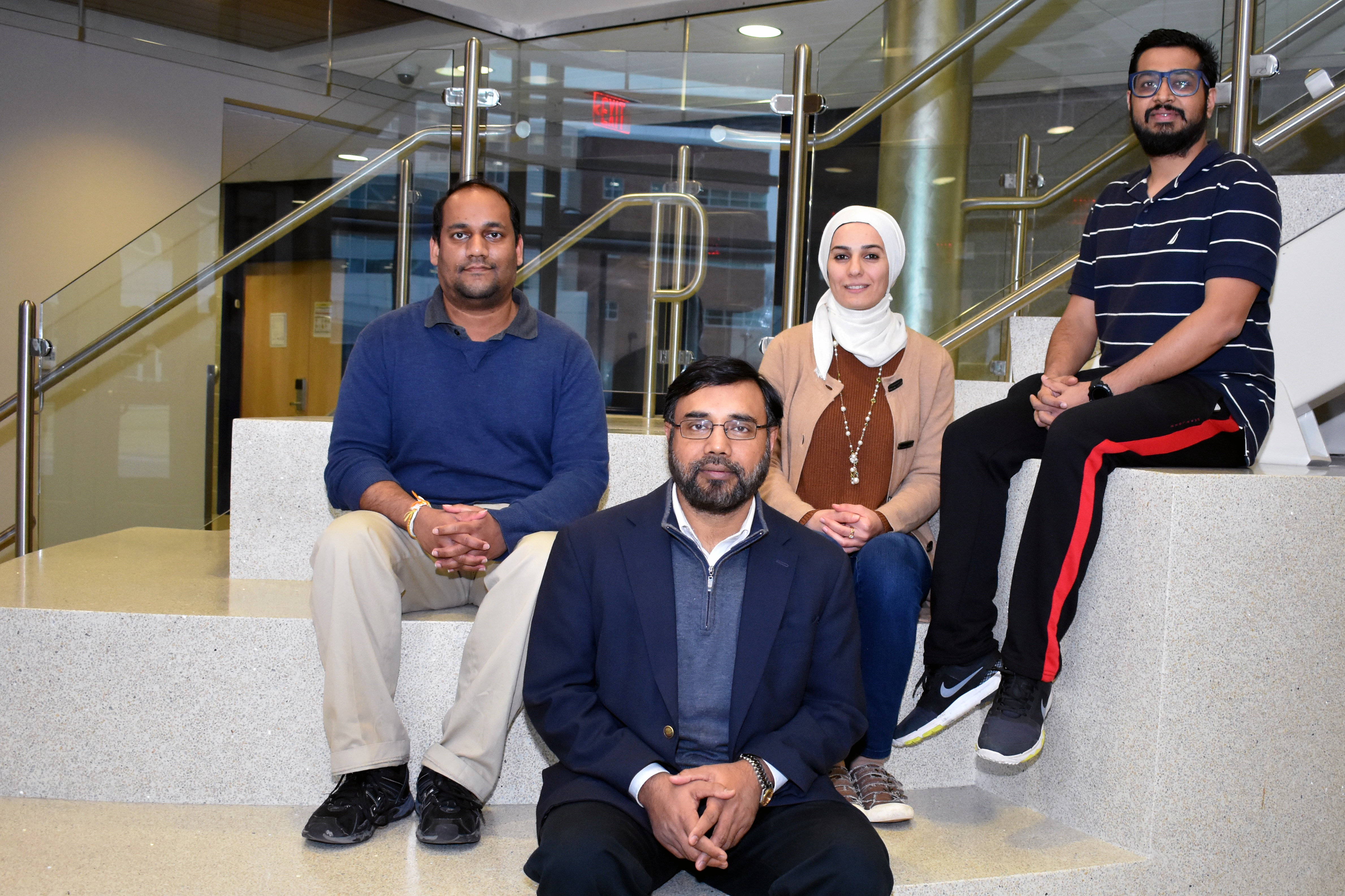
 Khan Iftekharuddin (foreground) and the Vision Lab team consisting of Lasitha Vidyaratne, Zeina Shboul and Mahbubul Alam.
By Keith Pierce
The MRI results are in, and when it comes to evaluating brain tumors using magnetic resonance imaging (MRI) scans, students from Old Dominion University's Vision Lab are among the best in the world.
ODU students placed first, out of 17 teams from around the globe, and ahead of the University Hospital of Bern (Switzerland), University of Castile-La Mancha (Ciudad Real, Spain) and the University of Virginia, in the Survival Prediction Category of the 2017 Multimodal Brain Tumor Segmentation (BraTS) Challenge, which was sponsored by the University of Pennsylvania.
Held in Quebec City, Quebec, the BraTS competition evaluated state-of-the-art methods for a process known as segmentation, the separation of brain tumor scans using computer vision in magnetic resonance imaging (MRI) and followed by prediction of survivability for patients.
Advised by Khan Iftekharuddin, associate dean for research for the College of Engineering and Technology and director of the ODU Vision Lab, the ODU team consisted of Ph.D. students Zeina Shboul, Lasitha Vidyaratne and Mahbubul Alam.
"This is a huge accomplishment for us," Iftekharuddin said. "This win placed the lab members, the Vision Lab and the University in the spotlight on the global stage. The Vision lab placed in the 3rd and 4th in BraTS challenges in prior years as well."
In the competition, student researchers are provided a large volume of training data consisting of MRI images captured using different scanners from multiple institutions.
The teams segment multiple abnormal tissues within the brain tumors from pre‐operative MRI scans and predict patient survival rates. Three categories of survival are considered: long survivors (less than 15 months), short survivors (less than 10 months), and mid survivors (between 10 and 15 months). Key factors such as the type of tumor, the mode in which the tumor is scanned and the stage of the disease affect segmentation.
The students are required to evaluate their segmentation and survival prediction performance and submit a short paper describing the results, as well as the segmentation method used. Their work is then evaluated and ranked against other competitors.
In the end, the ODU team placed first in the survival task for their paper entitled: Glioblastoma and Survival Prediction.
"Glioblastoma is categorized as a stage IV brain cancer," Iftekharuddin explained. "Our team in the Vision Lab is developing novel computational modeling and machine learning methods for segmentation of the tumors and, more importantly, survival prediction for patients affected by these lethal brain tumors. Such models will guide clinicians and practitioners in charting the course of action for these patients."
The BraTS challenge is organized as part of the Medical Image Computing and Computer-Assisted Intervention (MICCAI) conference, by the Section for Biomedical Image Analysis (SBIA), part of the Center for Biomedical Image Computing and Analytics (CBICA) in the Perelman School of Medicine at the University of Pennsylvania.

