Xu Research Group Strikes Gold with Nanoprobes
April 26, 2013
 Professor Nancy Xu
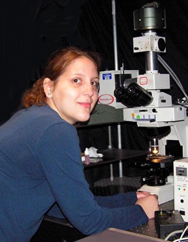
 Lauren Browning, doctoral student and Xu Group Researcher
 Tao Huang, an author of latest Xu Group research article
Medical diagnoses and therapies are expected to get big boosts from tiny nanoparticles, and groundwork for these advances is being accomplished today by the research group of X. Nancy Xu at Old Dominion University.
The group's latest research in nanobiotechnology, which has turned up fascinating details about nanoparticles made of gold, is reported this month in the special issue, "Molecular-, Nano- and Micro-Devices for Real-Time In Vivo Sensing," of the interdisciplinary journal Interface Focus from Royal Society Publishing in the United Kingdom. Titled "Real-time in Vivo Imaging of Size-dependent Transport and Toxicity of Gold Nanoparticles in Zebrafish Embryos Using Single Nanoparticle Plasmonic Spectroscopy," the article was authored by Lauren Browning, a doctoral student with the Xu group, Tao Huang, a research scientist who worked with Xu for several years, and Xu, professor of chemistry and biochemistry.
New experiments by the group show that relatively large gold nanoparticles - around 90 billionths of a meter in size - can penetrate live zebrafish embryos, disperse inside the organisms without being ejected and cause little to no damage. Previous journal articles by Xu and her associates have reported biotoxicity only slightly less promising for gold nanoparticles of less than 15 billionths of a meter. Silver nanoparticles, on the other hand, seem to have biotoxicity not detected in gold, according to the researchers.
Medical advances are anticipated if nanoparticles can penetrate human cells in order to act as biosensors or to deliver therapeutic drugs with pinpoint accuracy. If, indeed, the relatively large gold nanoparticles can penetrate cells without killing them, their size could be a boon for both imaging and drug delivery.
"This study offers new tools and new insights for the study of transport and toxicity of gold nanoparticles in vivo in real time at single nanoparticle resolution, which are essential to the rational design of biocompatible nanoparticles for a wide variety of biomedical applications, including in vivo imaging, drug delivery and implant devices," the authors write.
For a decade now, Xu has looked into a possible "stealth" quality for single-nanoparticle probes of living cells. She has been studying means by which nanoparticles can penetrate cells and accomplish their mission without harming the cells or being ejected by an efflux pumping mechanism that utilizes membrane transporters. This mechanism naturally targets foreign objects for ejection from cells.
Xu has found ways to synthesize and purify silver and gold nanoparticles that will stay stable - one size, or monodisperse - over an extended period. She and her colleagues have also reported breakthroughs in the way they image and characterize nanoparticles using dark-field optical microscopy and spectroscopy.
Being able to monitor nanoprobes in vivo - inside a living organism - is preferable, Xu said, to studies of nanotoxicity and other similar research done in vitro - on isolated cells in laboratory vessels. She said a more complete picture of biological processes is available from the single-nanoparticle probes in vivo.
But tracking single nanoparticles inside a living cell or embryo even for a split second is difficult, and the tracking is much more difficult over the 120 hours that Xu and her colleagues have been able to "watch" nanoprobes in a zebrafish embryo. Their latest imaging technique, which is reported in the Interface Focus article, involves localized surface plasmon resonance (LSPR) spectroscopy augmented by multi-spectral imaging system (MSIS) capability. The latter allows the researchers to simultaneously acquire LSPR spectra of a massive number of single nanoparticles, keeping tabs on the fate of the particles after they passively penetrate eggs. (The particles are suspended with the eggs in a liquid bath when this happens.) After a few days, the fertilized eggs have developed into embryos, and the researchers found that the particles typically became embedded throughout the fully developed organisms.
The great majority of the embryos - about 96 percent - developed normally with the 90-nanometer gold particles inside, showing no evidence of toxicity in the particles. This shows slightly better biocompatibility in the larger particles compared with the 12-nanometer particles the group had tested earlier. And the biocompatibility in the 90-nanometer gold particles turned out to be much better than that of the silver particles of about the same size.
These findings extend a long string of nanobiotechnology advances reported by the Xu group. Much of the work has been supported by grants totally more than $3 million from the National Science Foundation and National Institutes of Health.
A year ago, Xu, Huang and Browning wrote a cover story in the journal Nanoscale about their development of a photostable optical nanoscopy technique, which they call PHOTON. This far-field optical microscopy technique, when used in concert with the Xu group's single-molecule nanoparticle optical biosensors (SMNOBS), has enabled the team to map individual molecules and their binding with receptors on live cells in real time. These molecules - called ligands - can influence the functions of the cells they bind with. Precise information about the distribution of receptors and their number on single live cells is needed for the creation of more effective disease therapies, especially in the design of more effective vaccines.
That was followed by another cover story in Nanoscale by the same three authors reporting that they have been able to use PHOTON and SMNOBS to follow signaling pathways from the exterior membrane to the interior of the cell, and with exceptionally sharp resolution. Of specific interest to them is a single-molecule ligand called tumor necrosis factor-alpha (TNF-Alpha), which is a biomarker (indicator) for diagnosis of an array of diseases, including cancer, heart disease, diabetes and autoimmune diseases. The Xu group first reported the development of single-molecule nanoparticle optical biosensors of TNF-Alpha in Journal of the American Chemical Society in 2008.
TNF-Alpha can trigger a cascade of functions and lead to the orderly death of a cell, which is called apoptosis. This ligand is especially important because it can fight cancer. Unregulated apoptosis, of course, is not good; for example, it can result in atrophied body parts. But on the other hand, tumors can grow, sometimes remarkably fast, where apoptosis is inhibited. So knowing how apoptosis is signaled - or is not signaled - and revealing exactly how a cascade has to flow in order to bring about this orderly cell death is of paramount interest to scientists searching for a variety of therapies for human diseases.
Not only has the Xu group's research advanced techniques for nanoparticle delivery of drugs, but it also has found that the nanoparticles alone, without a drug payload - particularly the large-size silver nanoparticles - may be used to medicine's advantage by affecting certain cellular functions. This means that the nanoparticles themselves could be used as nanomedicines, say, to kill cancer cells, as well as serving as probes to illustrate molecular pathways about how cancer occurs and explore more effective therapy.
The Xu group has presented its work this year at the Association for the Advancement of Science annual meeting in Boston, at the Pittcon Laboratory Science Equipment Conference in Philadelphia and as a keynote at the Frontiers of Nano and Bioanalytical Chemistry meeting in New York City.

